Radiologists are facing barriers hindering AI adoption in clinical practice

Workflow integration
67% radiologists highlight workflow mapping and implementation as a barrier.1

Lack of trust
64 out of 100 CE-marked AI applications and products have no peer-reviewed evidence for their efficacy.2

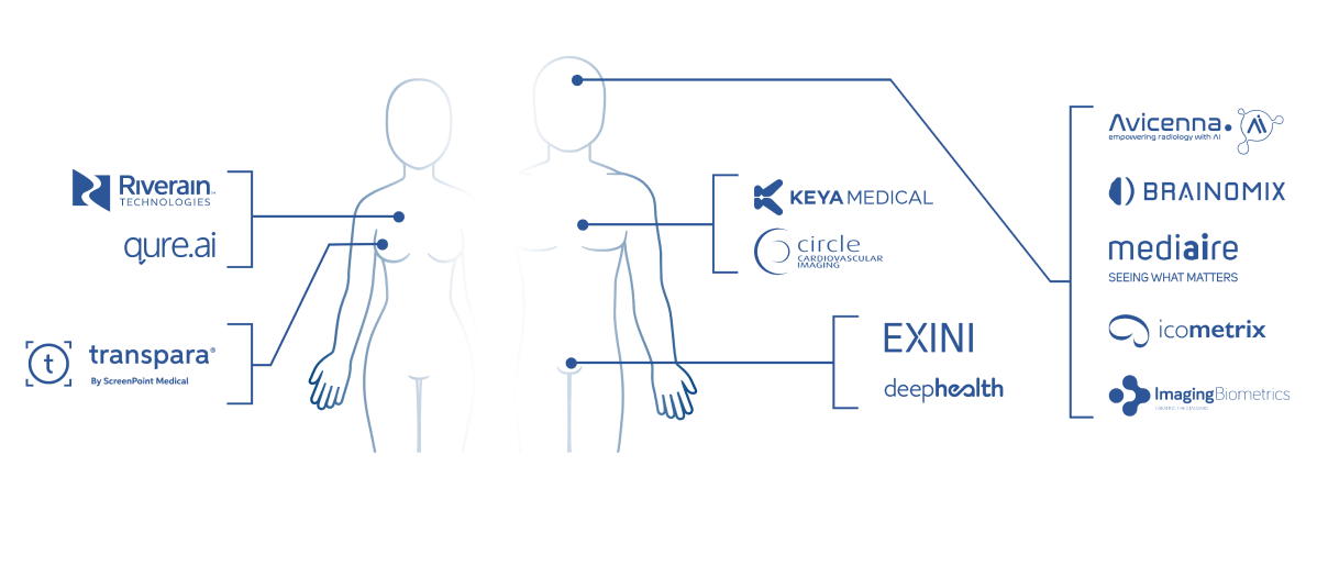
Rise of AI applications
More than 200 AI Radiology apps with CE certification in the EU.3

Rise of AI applications
More than 200 AI Radiology apps with CE certification in the EU.3

Cloud-hosted platform that provides access to AI applications

Access to all installed applications through a single common user interface

Zero-footprint CalanticTM Viewer integrated into the PACS viewport
Why Radiologists Trust Calantic

From the radiological-clinical perspective, at Instituts Guirado we are convinced that with the Calantic platform, there will be a before and after for lung nodules diagnosis and follow-up.”
Dr. Jorge Salmerón
Medical Director at Instituts Guirado

Delighted that Calantic joins us in our effort to provide the best possible state of the art imaging to our patients.”
Filip Van Grimberge
Head of Department Radiology AZ Turnhout


