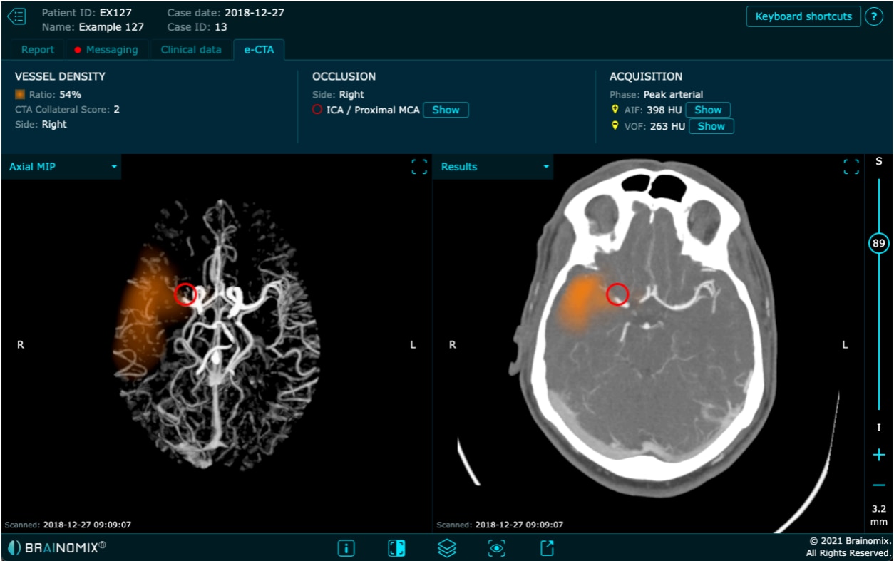
e-CTA is a software medical device for processing brain CT Angiography (CTA) scans for stroke patients and detect large vessel occlusions in brain CTA images. The software will attempt to find occlusions in the Internal Carotid Artery - Terminus (ICA-T), Middle Cerebral Artery, M1 Segment and Proximal Middle Cerebral Artery, M2 Segment arteries.1

Images shown for illustrative purposes only.
Brainomix 360 is a comprehensive platform designed to support clinicians and their imaging-based treatment decisions at all points across the stroke pathway, from simple imaging to more advanced imaging.
The Brainomix 360 Mobile App allows clinicians to quickly and securely access, preview, and share images and patient data across a network, send messages, make calls, and enhance collaboration for patient care - all designed to optimize workflow, facilitating faster transfer and treatment decisions.2

- Slice thickness: 1mm maximum slice thickness
- Acquisition volume: Full brain acquisition (e.g. Arch-to-vertex in a single sequence, or cerebrum sequence)
- Reconstruction method: Filtered back projection (FBP) with a moderate filter kernel (i.e. no excessive sharpening or smoothing)
- Bolus timing: Peak arterial or equilibrium phase (good acquisition timing) preferred
- Vessel collateral density
- Occlusion
- Acquisition

Currently not made available with Calantic Viewer. Separately distributed by Bayer.
EU risk class and CE marking
e-CTA has CE marking and risk class IIa.
Reimbursement status
Not reimbursed.
Contraindications
- e-CTA is not suitable for use on brain scans containing evidence of hemorrhage (intracranial, subarachnoid, etc.).
- e-CTA is not suitable for use on patients with posterior circulation stroke.
- e-CTA is not suitable for use on images that contain coils, shunts, embolization or movement artifacts.
- e-CTA is not suitable for use on brain scans displaying neurological pathologies other than acute ischaemic stroke, such as tumours or abscesses.
- e-CTA is not intended to be used for analyzing CTA images in intracranial vascular pathologies such as arterial aneurysms, arteriovenous malformations or venous thrombosis.
Target Population
Adults
Limitations
- Results generated by e-CTA should not be used as a standalone diagnosis.
- e-CTA should only be used by a competent healthcare professional with an understanding of brain CTA image interpretation, as an aid to image interpretation for estimation of collateral status.
- e-CTA may not correctly identify collaterals in every scan, and may generate false positives or false negatives. The users should use their own expertise in CTA image analysis to verify that the software’s output is correct.
- The user of e-CTA should monitor the image quality used for automated analysis in general, e.g. concerning motion artifacts.
- The user should also monitor the circumstances of contrast injection, which is also mandatory for medical reasons.
- e-CTA has only been validated in adult populations. Safety and effectiveness in pediatric cases has not been established.


Discover Brainomix e-CTA
Now available as part of Calantic’s Neuro Service Line. Contact us today to learn more.


