- MR
- CT
- CAD
- Cardiac
CAD and the Challenges of CCTA analysis
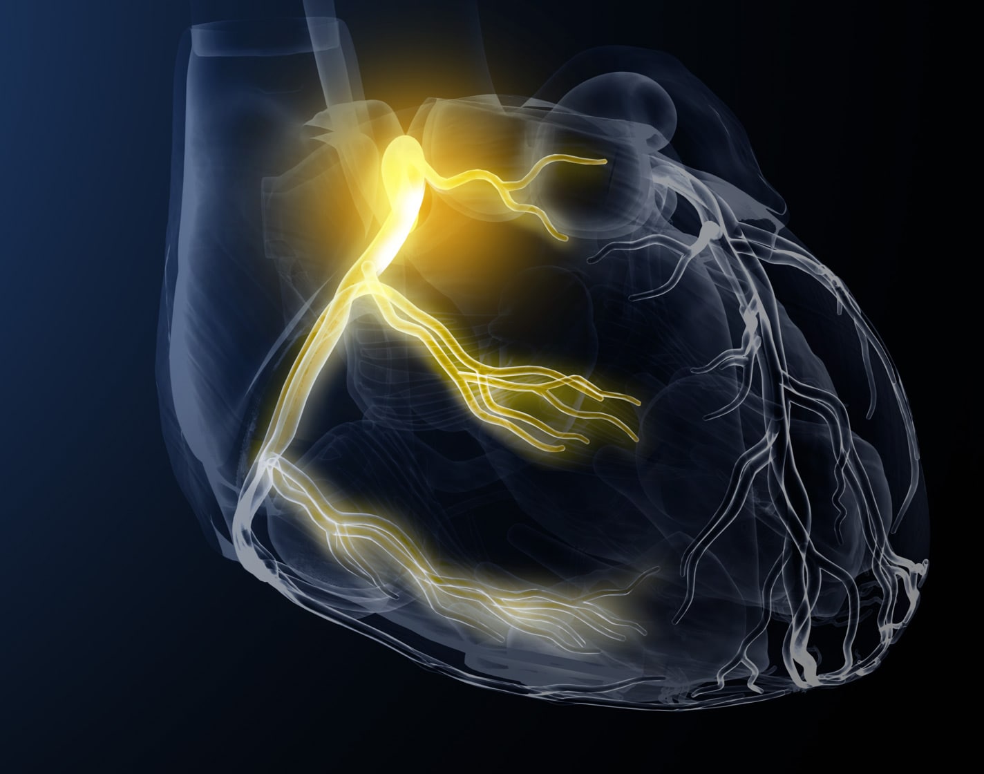
Coronary Artery Disease (CAD) is a known cause of significant cardiovascular events, accounting for more than 50% of the deaths in western countries2
Accurate interpretation of coronary computed tomography angiography (CCTA) is a labor-intensive and expertise-driven endeavor, as inexperienced readers may inadvertently overestimate stenosis severity3
Supporting cardiac MR & CT imaging analysis
cvi42 is intended to be used for viewing, post-processing, as well as for qualitative and quantitative evaluation of cardiovascular magnetic resonance (MR) images and cardiovascular computed tomography (CT) images in a Digital Imaging and Communications in Medicine (DICOM) Standard format.
It enables:
- Supporting clinical diagnostics by qualitative analysis of cardiac MR & CT images using display functionality such as panning, windowing, zooming, navigation through series/slices and phases, 3D reconstruction of images including multiplanar reconstructions of the images.
- Supporting clinical diagnostics by quantitative measurement of the heart and adjacent vessels in cardiac MR & CT images*, specifically signal intensity, distance, area, volume and mass.
- Supporting clinical diagnostics by using area and volume measurements for measuring LV function and derived parameters cardiac output and cardiac index in long axis and short axis cardiac MR & CT images.
- Flow quantification based on velocity encoded cardiac MR images including 2D and 4D flow analysis.
- Tissue characterization of cardiac MR Images.**
- Perfusion analysis of cardiac MR Images.**
- Strain analysis of cardiac MR images.**
- Supporting clinical diagnostics of cardiac CT images including quantitative measurements of calcified plaques in the coronary arteries (calcium scoring), specifically Agatston and volume and mass calcium scores, evaluation of heart structures including coronary arteries as well as aortic and mitral values.
- Evaluating CT and MR images of blood vessels. Combining digital image processing and visualization tools such as multiplanar reconstruction (MPR), thin/thick maximum intensity projection (MIP), inverted MIP thin/thick, volume rendering technique (VRT), curved planner reformation (CPR), processing tools such as bone removal (based on both single energy and dual energy) table removal and evaluation tools (vessel centerline calculation, lumen calculation, stenosis calculation) and reporting tools (lesion location, lesion characteristics) and key images.
- The software package is designed to support the physician in confirming the presence or absence of physician identified lesions in blood vessels and evaluation, documentation and follow up of any such lesions.
*Quantitative analysis ls dependent on the quality and correctness of the image source data
**Tissue, Perfusion, & Strain ( 3D & RV) features are not available for clinical use in the USA. These modules are to be used for research purposes only, and not for primary diagnostics and direct patient care.
Diagnostic Accuracy. Efficiency. Patient Care.

Viewer
- Multiple image synchronization options.
- Multiple contouring and measurement tools.
- Compare baseline and follow-up scans.
Patient Data
- Review and edit study data.
- Create case review presentations.

Reporting
- Results and reference values can be Imported into reports by the radiologist.
- Create custom report templates.
- HL7 compatible.
1 Circle Cardiovascular Imaging. cvi42 User Manual Version 6.0, January 2024
2 Shreya 0, Zamora DI, Patel GS, Grossmann I, Rodriguez K, Soni M, Joshi PK, Patel SC, Sange I. Coronary Ar tery Calcium Score -A Reliable Indicator of Coronary Artery Disease? Cureus. 2021 Dec 3;13(12J:e20149. doi: 10.7759lureus.20149. PMID: 35003981; PMCIO: PMC8723785.
3 Meng Q, Yu P, Yin S, Li X, Chang Y, Xu W, Wu C, Xu N, Zhang H, Wang Y, Shen H, Zhang R, Zhang Q. Coronary computed tomography angiography analysis using artificial intelligence for stenosis quantification and stent segmentation: a multlcenter study. Quant Imaging Med Surg. 2023 Oct 1;13(10):6876-6886. doi: 10.21037/ qlms- 23-423. Epub 2023 Sep 15. PMIO: 37869330; PMCIO: PMC10585569.
4 Halipoglu, Suzan, et al. *Performance or art ificial intelligence for biventrlcular cardiovascular magnetic resonance volumetric analysls in the clinical selling." The International Journal of Cardiovascular Imaging 38.11 (2022): 2413-2424.
5 Urmeneta Ulloa, Javier, et al. "Comparative Cardiac Magnetic Resonance-Based Feature Tracking and Deep-Learning Strain Assessment in Patients Hospitalized for Acute Myocardltis.* Journal oi Clinical Medicine 12.3 (2023): 1113.
6 Ayton, Sarah L., et al. "The lnterfield Strength Agreement of Left Ventricular Strain Measurements at 1.5 T and 3 T Using Cardiac MRI Feature Tracking." Journal of Magnetic Resonance Imaging 57.4 (2023): 1250-1 261.
7 Militaru, Sebastian, et al. "Mul tivendor comparison of global and regional 20 cardiovascular magnetic resonance feature tracking st rains vs tissue tagging at 3T." Journal of Cardiovascular Magnetic Resonance 23 (2021): 1-
Currently not available within the Calantic Viewer.


